Hammerhead
Location
Room 0832. Veritas Science Center (Former NRB)
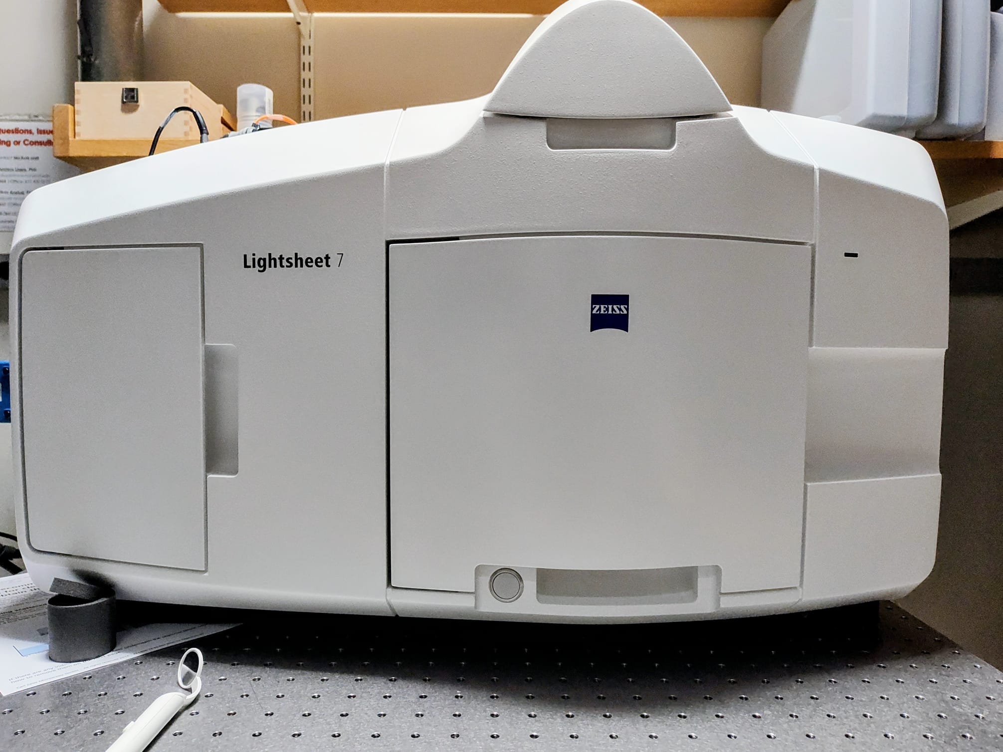
Zeiss Lightsheet 7
Multi-view imaging in a variety of samples ranging from live organisms and organoids to fixed samples that have been optically cleared by both water-based (expansion, CLARITY, CUBIC, Scale) and solvent-based (iDisco)methods.
NIH S10 Acknowledgment
The Zeiss LS7 lightsheet micrsoscoscope was funded through a NIH-S10 Instrumentation grant. NIH requires us to report every NIH grant/project that benefits from using this instrument. Additionally, NIH requires all researchers to acknowledge this grant in any publication using the instrument (Zeiss LS7). Please add the following grant to the funding sources/acknowledgments in any publication or grant submitted: Please use this grant number 1S10OD036362
Hardware
Zeiss Lightsheet 7 with double side illumination
For cleared and live samples (37°C incubator and CO2)
Objectives
Lightsheet Illumination objectives
- 5x / NA-o.1 (for lower res illumination)
- 10x / NA - 0.2 (for higher res illumination)
Detection objectives
- 5x/0.16 Plan Neofluar (for overview images of large samples)
- 10x/0.5 Plan Apochromat Water dipping (for live cell imaging of large samples or water-based-cleared samples)
- 20x/1.0 Plan Apochromat Water dipping (for high resolution live cell imaging)
- 20x/1.0 Plan Apochromat with correction collar (for clearing solutions with RI of 1.45)
- 20x/1.0 Plan Neofluar with correction collar (for clearing solutions with RI of 1.53)
Filters and dichroics
- Quad dichroic 405/488/561/638
- emission filter (dual emission)
- DAPI (BP420-470) + GFP(BP505-545)
- DAPI (BP420-470) + CY3 (BP575-615)
- GFP (BP505-545) + mCherry, DRAQ5 (LP585)
- GFP (BP505-545) + DRAQ5 (LP660)
- Cy3 (BP575-615) + DRAQ5 (LP660)
Cameras
- 2x PCO Edge 4.2 sCMOS - (6.5um pixel size)
Lasers
- 405
- 488
- 561
- 638